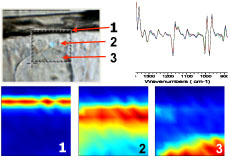
WHAT IS MIRAS ?
Infrared (IR) spectroscopy is undoubtedly the most widely used characterisation technique in the world due to its wide versatility and the comprehensive information it can provide for the study of molecules. The combination of IR spectroscopy with microscopy employing a synchrotron as the IR source provides an extremely powerful tool with wide applicability. In recent years the growth of SIRMS (or MIRAS) has been spectacular precisely due to its great success in diverse fields, ranging from biology and biomedicine, forensics and industrial analysis, archeometry and cultural heritage, to polymer and ceramic materials science, geology and astrophysics, amongst others. Currently there are over 20 active synchrotron IR beamlines, and the construction of new beamlines for IR spectroscopy and microspectroscopy are projected in almost all the world's synchrotron facilities.
SHORT HISTORY OF MIRAS
A low divergence, diffraction-limited infrared point source with a very high photon flux per source area and solid angle, i.e. high brightness, two to three orders of magnitude greater than that of a conventional blackbody source1,2, can be perfectly coupled to a microscope since all of the flux can be efficiently concentrated in a very small area, between 5 – 10 μm. This ideal IR source is provided by a synchrotron. It has been shown that in the mid IR region (< 25 μm) a factor of around 1000x improvement in brightness and, as a consequence, a spectacular increase in the signal-to-noise ratio.
 The use of a synchrotron (s2) as an IR source is known since 1982 when it was experimentally demonstrated at Brookhaven National Laboratory , USA.3 The first synchrotron IR microspectroscopy (SIRMS) beamline was built at Brookhaven National Laboratory in 1993.4 Since then, the technique has rapidly expanded, and around 15 SIRMS beamlines are currently operating worldwide, and many more are projected in the coming years which will greatly improve the accessibility to this powerful, cost effective analytical tool.
REFERENCIAS
- W.D. Duncan, G.P. Williams, Appl. Opt. 22 (1983) 2914
- G.L. Carr, P. Dumas, C.J. Hirschmugl, G.P. Williams, Il Nuovo Cim. 20 (1998) 375
- G.P. Williams, Nucl. Instr. and Meth. 195 (1982) 383
- J.A. Reffner, P.A. Martoglio, G.P. Williams, Rev. Sci. Instrum. 66 (1995) 1298
TYPICAL CHARACTERISTICS OF A BEAMLINE
There are many active IR beamlines both in Europe and the rest of the world. Links to many are included at the bottom of this page. In the following table some typical figures and characteristics of a modern IR beamline are provided*:
| Typical specifications | |
Source |
Bending Magnet (BM) or Edge radiation (ER) or both |
Energy range |
0.4 µm to 100 µm |
Flux at 1st optical element |
> 1 e+14 Phot/s/0.1%bw at 10 µm for 500 mA stored current |
Energy resolution |
Depends on interferometric system. Can vary typically between 0.5 cm-1 (standard) and 0.0007 cm-1 (high resolution) |
Beam size at sample (microscopy) |
Between 5x5 µm2 and 15x15 µm2 |
Flux at spectrometer |
> 2 e+14 Phot/s/0.1%bw at 10 µm before entrance |
Detectors |
Mid, near and far-IR detectors. Imaging systems (FPA) |
| * The actual values vary and depend on the design characteristics of the synchrotron. | |
WHAT IS "MIRAS" USEFUL FOR?
Follow these links for examples of experiments with MIRAS (SIRMS) .
- INFRARED IMAGING from lightsources.org
- PUBLICATION LIST from ALS-Berkeley Laboratory, USA
- ABSTRACTS from the lectures at the MIRAS-2008 Workshop
- RESEARCH of the Miller group from NSLS-Brookhaven National Laboratory, USA
- PUBLICATION from the U2A beamline, NSLS on High Pressure and Microscopy
MIRAS IN THE WORLD
This section presents links to some synchrotrons with IR beamlines:
 ALS, LBL, Berkeley, CA, USA
- ANKA, Karlsruhe, Germany
- AS, Melbourne, Australia
- BESSY, Berlin, Germany
- CLS, Saskatoon, Canada
- DAFNE, Frascati, Italy
- DIAMOND - Didcot, UK
- ELETTRA, Trieste, Italy
- MAX Lab, Lund, Sweden
- NSLS, BNL, Upton, NY, USA
- NSRRC, Hsinchu, Taiwan
- PAL, Pohang , Korea
- SLS, Villigen, Switzerland
- SOLEIL, Orsay, France
- SPring-8, Harima, Japan
- SRC, Stoughton, WI, USA
A more recent update of IR beamlines can be found in the following review.